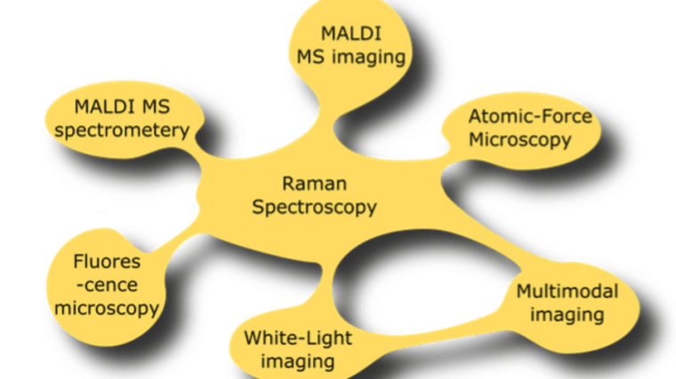
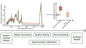
Fig 3

Photonic Data Science

Research group Prof. Dr. Thomas Bocklitz
Fig 3
Graphic: IPHT

Head of the group

Thomas Bocklitz, Univ.-Prof. Dr
Head
Professorship of Photonic Data Science
Prof. Dr. Thomas Bocklitz
Image: Prof. Dr. Thomas Bocklitz
JenTower
Leutragraben 1
07743 Jena Google Maps site planExternal link

Scientific Profile

We explore the entire data life cycle of photonic data from generation to the data analysis and to data archiving. Following a holistic approach, we investigate procedures for experiment and sample size planning as well as data pretreatment and combine these procedures with chemometric procedures, model transfer methods and artificial intelligence methods in a data pipeline. In this way, data from various photonic processes can be used for analysis, diagnostics and therapy in medicine, life science, environmental sciences and pharmacy. The data pipeline are implemented in software components and are tested directly in the applicative environment, e.g. in clinical studies. Further focal points are data fusion of different heterogeneous data sources, the simulation of different measurement procedures in order to optimize correction procedures, methods for the interpretation of analysis models and the construction of data infrastructures for different photonic measurement data, which ensure the FAIR principles.

Research Topics

  • Machine learning for photonic image data

    Fig 1

    Graphic: IPHT
  • Chemometrics / machine learning for spectral data
  • Correlation of different measurement methods and data fusion

Areas of application

  • Bio-medical diagnostics using spectral measurement methods and imaging techniques
  • Extraction of higher information from photonic measurement data
  • Simulation- und data-driven correction of photonic data
  • Guarantee of FAIR principles for photonic data

Staff

  1. Adhikari, Mou PhD Student Professorship of Photonic Data Science

    JenTower, Room 15S04
    Leutragraben 1
    07743 Jena

  2. Amiri, Mahmoud Professorship of Photonic Data Science

    JenTower
    Leutragraben 1
    07743 Jena

  3. Azam, Kazi Sultana Farhana PhD student Professorship of Photonic Data Science

    JenTower, Room 15S04
    Leutragraben 1
    07743 Jena

  4. Contreras Duarte, Jhonatan PostDoc Professorship of Photonic Data Science

    JenTower, Room 15S02
    Leutragraben 1
    07743 Jena

  5. Corbetta, Elena PhD student Professorship of Photonic Data Science

    JenTower, Room 15S05
    Leutragraben 1
    07743 Jena

  6. Darzi, Fatemehzahra PhD student Professorship of Photonic Data Science

    JenTower, Room 15S03
    Leutragraben 1
    07743 Jena

  7. Dehbozorgi, Pegah PhD Student Professorship of Photonic Data Science

    JenTower, Room 15S03
    Leutragraben 1
    07743 Jena

  8. Kamran, Jawad Professorship of Photonic Data Science

    JenTower, Room 15S07
    Leutragraben 1
    07743 Jena

  9. Mokari, Azadeh PhD Student Professorship of Photonic Data Science

    JenTower, Room 15S01
    Leutragraben 1
    07743 Jena

  10. Morgunov, Volodymyr, Dr PostDoc Professorship of Photonic Data Science

    JenTower, Room 15S07
    Leutragraben 1
    07743 Jena

    Dr. Volodymyr Morgunov
    Image: Volodymyr Morgunov
  11. Mostafapourghasrodashti, Sara PhD student Professorship of Photonic Data Science
  12. Ryabchykov, Oleg, Dr PostDoc Professorship of Photonic Data Science

    JenTower, Room 15S02
    Leutragraben 1
    07743 Jena

  13. Stefaniuk, Nazar PostDoc Professorship of Photonic Data Science

    JenTower
    Leutragraben 1
    07743 Jena

  14. Thamm, Sophie, Dr Coordinator LPI Professorship of Photonic Data Science

    JenTower, Room 15S08
    Leutragraben 1
    07743 Jena

  15. Vulchi, Ravi Teja PhD Student Professorship of Photonic Data Science

    JenTower
    Leutragraben 1
    07743 Jena

  16. Yogita, Yogita PhD student Professorship of Photonic Data Science

    JenTower, Room 15S01
    Leutragraben 1
    07743 Jena

54 Publikationen filtern

Die Publikationen filtern

Highlighted authors are members of the University of Jena.

  1. Explainable artificial intelligence for spectroscopy data: a review

    Year of publicationPublished in:Pflügers Archiv : European journal of physiology J. Contreras, T. Bocklitz
    Explainable artificial intelligence (XAI) has gained significant attention in various domains, including natural and medical image analysis. However, its application in spectroscopy remains relatively unexplored. This systematic review aims to fill this gap by providing a comprehensive overview of the current landscape of XAI in spectroscopy and identifying potential benefits and challenges associated with its implementation. Following the PRISMA guideline 2020, we conducted a systematic search across major journal databases, resulting in 259 initial search results. After removing duplicates and applying inclusion and exclusion criteria, 21 scientific studies were included in this review. Notably, most of the studies focused on using XAI methods for spectral data analysis, emphasizing identifying significant spectral bands rather than specific intensity peaks. Among the most utilized AI techniques were SHapley Additive exPlanations (SHAP), masking methods inspired by Local Interpretable Model-agnostic Explanations (LIME), and Class Activation Mapping (CAM). These methods were favored due to their model-agnostic nature and ease of use, enabling interpretable explanations without modifying the original models. Future research should propose new methods and explore the adaptation of other XAI employed in other domains to better suit the unique characteristics of spectroscopic data.
    University Bibliography Jena:
    fsu_mods_00016079External link
  2. Machine Learning-Based Estimation of Experimental Artifacts and Image Quality in Fluorescence Microscopy

    Year of publicationPublished in:Advanced Intelligent Systems E. Corbetta, T. Bocklitz
    Reliable characterization of image data is fundamental for imaging applications, FAIR data management, and an objective evaluation of image acquisition, processing, and analysis steps in an image-based investigation of biological samples. Image quality assessment (IQA) often relies on human visual perception, which is not objective, or reference ground truth images, which are not often available. This study presents a method for a comprehensive IQA of microscopic images, which solves these issues by employing a set of reference-free metrics that estimate the presence of experimental artifacts. The metrics are jointly validated on a semisynthetic dataset and are tested on experimental images. Finally, the metrics are employed in a machine learning model, demonstrating their effectiveness for automatic artifact classification through multimarker IQA. This work provides a reliable reference-free method for IQA in optical microscopy, which can be integrated into the experimental workflow and tuned to address specific artifact detection tasks.
    University Bibliography Jena:
    fsu_mods_00018351External link
  3. Enhancing prediction stability and performance in LIBS analysis using custom CNN architectures

    Year of publicationPublished in:Talanta: the international journal of pure and applied analytical chemistry P. Dehbozorgi, L. Duponchel, V. Motto-Ros, T. Bocklitz
    LIBS-based analysis has experienced an ever-increasing interest in recent years as a well-suited technique for chemical analysis tasks relying on elemental fingerprinting. This method stands out for its ability to offer rapid, simultaneous multi-element analysis with the advantage of portability. In the context of this research, our aim is to bridge the gap between the analysis of simulated and real data to better account for variations in plasma temperature and electron density, which are typically not considered in LIBS analysis. To achieve this, we employ two distinct methodologies, PLS and CNNs, to construct predictive models for predicting the concentration of the 24 elements within each LIBS spectrum. The initial phase of our investigation concentrates on the training and testing of these models using simulated LIBS data, with results evaluated through RMSEP values. The IQR and median RMSEP values for all the elements demonstrate that CNNs consistently achieved values below 0.01, while PLS results ranged from 0.01 to 0.05, highlighting the superior stability and predictive accuracy of CNNs model. In the next phase, we applied the pre-trained models to analyze the real LIBS spectra, consistently identifying Aluminum (Al), Iron (Fe), and Silicon (Si) as having the highest predicted concentrations. The overall predicted values were approximately 0.5 for Al, 0.6 for Si, and 0.04 for Fe. In the third phase, deliberate adjustments are made to the training parameters and architecture of the proposed CNNs model to force the network to emphasize specific elements, prioritizing them over other components present in each real LIBS spectrum. The generation of the three modified versions of the initially proposed CNNs allows us to explore the impact of regularization, sample weighting, and a customized loss function on prediction outcomes. Some elements emerge during the prediction phase, with Calcium (Ca), Magnesium (Mg), Zinc (Zn), Titanium (Ti), and Gallium (Ga) exhibiting more pronounced patterns.
    University Bibliography Jena:
    fsu_mods_00018242External link
  4. 3D Hyperspectral Data Analysis with Spatially Aware Deep Learning for Diagnostic Applications

    Year of publicationPublished in:Analytical Chemistry R. Luo, S. Guo, J. Hniopek, T. Bocklitz
    Nowadays, with the rise of artificial intelligence (AI), deep learning algorithms play an increasingly important role in various traditional fields of research. Recently, these algorithms have already spread into data analysis for Raman spectroscopy. However, most current methods only use 1-dimensional (1D) spectral data classification, instead of considering any neighboring information in space. Despite some successes, this type of methods wastes the 3-dimensional (3D) structure of Raman hyperspectral scans. Therefore, to investigate the feasibility of preserving the spatial information on Raman spectroscopy for data analysis, spatially aware deep learning algorithms were applied into a colorectal tissue data set with 3D Raman hyperspectral scans. This data set contains Raman spectra from normal, hyperplasia, adenoma, carcinoma tissues as well as artifacts. First, a modified version of 3D U-Net was utilized for segmentation; second, another convolutional neural network (CNN) using 3D Raman patches was utilized for pixel-wise classification. Both methods were compared with the conventional 1D CNN method, which worked as baseline. Based on the results of both epithelial tissue detection and colorectal cancer detection, it is shown that using spatially neighboring information on 3D Raman scans can increase the performance of deep learning models, although it might also increase the complexity of network training. Apart from the colorectal tissue data set, experiments were also conducted on a cholangiocarcinoma data set for generalizability verification. The findings in this study can also be potentially applied into future tasks regarding spectroscopic data analysis, especially for improving model performance in a spatially aware way.
    University Bibliography Jena:
    fsu_mods_00024120External link
  5. Correction to: SERS-Driven Ceftriaxone Detection in Blood Plasma: A Protein Precipitation Approach (Chemosensors, (2024), 12, 10, (213), 10.3390/chemosensors12100213)

    Year of publicationPublished in:Chemosensors A. Dwivedi, O. Ryabchykov, C. Liu, E. Farnesi, M. Schmidt, T. Bocklitz, J. Popp, D. Cialla-May
    The blood-based samples for the original publication [1] were purchased from the Institut für Klinische Transfusionsmedizin Jena gGmbH (Jena, Germany). Thus, no ethics approval and informed consent are needed. However, the Institutional Review Board Statement of the paper should not be “Not applicable”, and it should be changed to a specific reason why the ethics approval can be waived. A correction has been made to Institutional Review Board Statement: Institutional Review Board Statement: The blood samples were purchased from the Institut für Klinische Transfusionsmedizin Jena gGmbH (Jena, Germany). All methods were performed in accordance with the relevant guidelines and regulations of the clinics of Universitätsklinikum Jena, thus the blood samples are exempt from the need for ethical approval. The authors state that the scientific conclusions are unaffected. This correction was approved by the Academic Editor. The original publication has also been updated.
    University Bibliography Jena:
    fsu_mods_00020927External link
  6. Bridging Spectral Gaps: Cross-Device Model Generalization in Blood-Based Infrared Spectroscopy

    Year of publicationStatusReview pendingPublished in:Analytical chemistry / publ. by the American Chemical Society. Ed. dir. Walter J. Murphy F. Nemeth, N. Leopold-Kerschbaumer, D. Debreceni, F. Fleischmann, K. Borbely, D. Mazurencu-Marinescu-Pele, T. Bocklitz, M. Žigman, K. Kepesidis
    This paper presents a solution to the challenge of cross-device model generalization in blood-based infrared spectroscopy. As infrared spectroscopy becomes increasingly popular for analyzing human blood, ensuring that machine learning models trained on one device can be effectively transferred to others is essential. However, variations in device characteristics often reduce model performance when applied across different devices. To address this issue, we propose a straightforward domain adaptation method based on data augmentation incorporating device-specific differences. By expanding the training data to include a broader range of nuances, our approach enhances the model’s ability to adapt to the unique characteristics of various devices. We validate the effectiveness of our method through experimental testing on two Fourier-Transform Infrared (FTIR) spectroscopy devices from different research laboratories, demonstrating improved prediction accuracy and reliability.
    University Bibliography Jena:
    fsu_mods_00024831External link
  7. Physics-informed neural networks for etaloning correction in Raman spectra using inverse modeling

    Year of publicationStatusReview pendingPublished in:Proceedings of SPIE - The International Society for Optical Engineering R. Vulchi, V. Morgunov, T. Bocklitz
  8. A comparative study of statistical, radiomics, and deep learning feature extraction techniques for medical image classification in optical and radiological modalities

    Year of publicationStatusReview pendingPublished in:Computers in biology and medicine : an international journal P. Dehbozorgi, O. Ryabchykov, T. Bocklitz
    Feature extraction in ML plays a crucial role in transforming raw data into a more meaningful and interpretable representation. In this study, we thoroughly examined a range of feature extraction techniques and assessed their impact on the binary classification models for medical images, utilizing a diverse and rich set of medical imaging modalities. Using H&E-stained, chest X-ray, and retina OCT images, we applied methods to extract statistical, radiomics, and deep features. These features were then used to develop PCA-LDA models as the employed classifier. We evaluated the models based on two decisive metrics: latency and performance. Latency measured the time taken for feature extraction and prediction, while mean sensitivity (balanced accuracy) characterizes the model performance. Our comparative study revealed that statistical and radiomics features were less effective for medical image classification, as they showed high latency and lower performance scores. In contrast, pre-trained DL networks performed efficiently, with high sensitivity and low latency. For H&E-stained images, the statistical feature extraction took about an hour and achieved 90.8 % sensitivity, while ResNet50 reduced processing time fourfold and increased sensitivity to 96.9 %. For chest X-rays, radiomics features were time-intensive with 92.2 % sensitivity, while ResNet50 improved sensitivity to 96 % with faster extraction time. For retina OCT images, radiomics yielded a sensitivity of 91 %, while DenseNet121 achieved 98.6 % sensitivity in 15 min. These findings underscore the superior performance of DL techniques over the statistical and radiomics features, highlighting their potential for real-world applications where accurate and rapid diagnostic decisions are essential.
    University Bibliography Jena:
    fsu_mods_00019907External link
  9. Long-term device stability for Raman spectroscopy

    Year of publicationStatusReview pendingPublished in:Analyst S. Guo, A. Ramoji, A. Pistiki, H. Yilmaz, U. Glaser, D. Vasquez-Pinzon, I. Schie, U. Neugebauer, A. Silge, J. Popp, T. Bocklitz
  10. MMIR: an open-source software for the registration of multimodal histological images

    Year of publicationPublished in:BMC Medical Informatics and Decision Making R. Escobar Díaz Guerrero, J. Oliveira, J. Popp, T. Bocklitz
    Background: Multimodal histology image registration is a process that transforms into a common coordinate system two or more images obtained from different microscopy modalities. The combination of information from various modalities can contribute to a comprehensive understanding of tissue specimens, aiding in more accurate diagnoses, and improved research insights. Multimodal image registration in histology samples presents a significant challenge due to the inherent differences in characteristics and the need for tailored optimization algorithms for each modality. Results: We developed MMIR a cloud-based system for multimodal histological image registration, which consists of three main modules: a project manager, an algorithm manager, and an image visualization system. Conclusion: Our software solution aims to simplify image registration tasks with a user-friendly approach. It facilitates effective algorithm management, responsive web interfaces, supports multi-resolution images, and facilitates batch image registration. Moreover, its adaptable architecture allows for the integration of custom algorithms, ensuring that it aligns with the specific requirements of each modality combination. Beyond image registration, our software enables the conversion of segmented annotations from one modality to another.
    University Bibliography Jena:
    fsu_mods_00011421External link
  11. Non-resonant background removal in broadband CARS microscopy using deep-learning algorithms

    Year of publicationPublished in:Scientific Reports F. Vernuccio, E. Broggio, S. Sorrentino, A. Bresci, R. Junjuri, M. Ventura, R. Vanna, T. Bocklitz, M. Bregonzio, G. Cerullo, H. Rigneault, D. Polli
    Broadband Coherent anti-Stokes Raman (BCARS) microscopy is an imaging technique that can acquire full Raman spectra (400–3200 cm−¹) of biological samples within a few milliseconds. However, the CARS signal suffers from an undesired non-resonant background (NRB), deriving from four-wave-mixing processes, which distorts the peak line shapes and reduces the chemical contrast. Traditionally, the NRB is removed using numerical algorithms that require expert users and knowledge of the NRB spectral profile. Recently, deep-learning models proved to be powerful tools for unsupervised automation and acceleration of NRB removal. Here, we thoroughly review the existing NRB removal deep-learning models (SpecNet, VECTOR, LSTM, Bi-LSTM) and present two novel architectures. The first one combines convolutional layers with Gated Recurrent Units (CNN + GRU); the second one is a Generative Adversarial Network (GAN) that trains an encoder-decoder network and an adversarial convolutional neural network. We also introduce an improved training dataset, generalized on different BCARS experimental configurations. We compare the performances of all these networks on test and experimental data, using them in the pipeline for spectral unmixing of BCARS images. Our analyses show that CNN + GRU and VECTOR are the networks giving the highest accuracy, GAN is the one that predicts the highest number of true positive peaks in experimental data, whereas GAN and VECTOR are the most suitable ones for real-time processing of BCARS images.
    University Bibliography Jena:
    fsu_mods_00017674External link
Pagination Page 1